How sport promotes blood flow to muscles
ETH professor Katrien De Bock and her team have found a certain type of blood vessel cell in muscles that multiplies particularly quickly during exercise and thus forms new blood vessels. This means that the researchers can now get to the bottom of circulatory disorders in muscles.


"The most common reason why surgeons in industrialized countries have to amputate someone's foot or leg is inadequate blood supply to muscles in diabetes patients," says Katrien De Bock. She is a professor of exercise and health at ETH Zurich and is working with her team to investigate how such circulatory disorders in muscles can be treated and how blood vessels are formed anew. It is well known that exercise and sport stimulate blood vessel formation. However, the molecular and cellular mechanisms by which this occurs are poorly understood. "If we understand these mechanisms, we can work toward specifically promoting blood supply to muscles in patients," says the ETH professor.
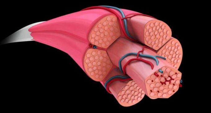
In mice and in cell culture of human cells, De Bock and her colleagues have now investigated how the fine blood vessel capillaries are formed in muscles of healthy individuals. They have targeted the vessel wall cells (endothelial cells) and discovered that there are two types of endothelial cells that differ with respect to a molecular marker called ATF4. Cells with low levels of ATF4 are mainly found in capillaries, which supply so-called white muscle fibers. And cells with a lot of ATF4 are mainly part of the blood vessels near red muscle fibers, the researchers found.
"Ready to go"
The scientists further showed that physical exercise stimulates endothelial cells with high ATF4 (i.e., those in red muscle fibers) to divide, resulting in the formation of new blood vessel capillaries. Cells with little ATF4, on the other hand, do not respond directly to exercise. "The endothelial cells with a lot of ATF4 are sort of on standby," De Bock says. ATF4 is a regulatory protein inside cells. Cells with this protein are ready to respond quickly to the appropriate stimulus: As soon as a person - or in our case a mouse - exercises, these cells take up more amino acids and invest in increased formation of DNA and proteins and in the rapid proliferation of cells. This ultimately leads to the formation of new vessels.
It is not yet known why these "ready to go" vascular wall cells are mainly located near the red muscle fibers. The researchers would like to investigate this next. In addition, the scientists would like to use the findings to develop therapies to stimulate the growth of muscular blood vessels in diabetics, patients with arterial occlusive diseases or with transplanted organs.
Source: ETH News